Special tests mean extra special eye care
Optical Coherence Tomographer (OCT)
Our OCT helps us better manage glaucoma and diseases of the retina because this technology allows the eye doctor to see the deep tissue layers in the eye. Similar to ultrasound, this diagnostic technique employs light rather than sound waves to achieve higher resolution pictures of the structural layers of the back of the eye. These high-definition images are the only way that they can actually see beneath the surface to the nerve fiber layers where damage occurs. Up until now, eye doctors had to use other tests to indicate damage in this critical area of sight. Common eye diseases such macular degeneration, diabetic retinopathy, and glaucoma are detected early by the OCT when the diseases can be more effectively treated.
ZEISS / Humphrey Visual Field Testing
Visual Field testing can help save vision because it is another test used to diagnose or rule out glaucoma and other neurological disorders that affect vision. This simple, but effective service has saved lives by detecting various medical conditions such as strokes, brain tumors, and other neurological defects.
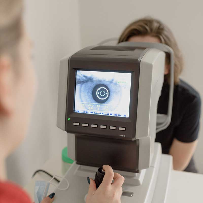
i.Terminal 2
Lens fitting plays a key role in maximizing visual comfort, as fitting errors can cause up to a 40% loss in lens performance. i.Terminal 2 captures and calculates your patient´s individual parameters with the click of a button and a precision of 0.1 mm which can result in a decreased complaint rate, reduced non-adapts and more relaxed vision for your patients.
Fundus Photography, or Retinal Digital Imaging
A high-definition digital image of the retinal area helps your eye doctor diagnose and manage eye diseases in the delicate retinal area. Damage to the delicate structures of the retina is often the first sign of systemic diseases such as MS, diabetes and others. The retina is the “window to the body” and routine retinal imaging can help your eye doctor monitor the changes in your eye health from year to year.
Dry Eye Treatment
Our tears contain three layers — oil (lipid) layer, water (aqueous) layer and a mucin layer. These three layers are responsible for keeping the eye surface lubricated. Our meibomian glands located along the rims of the eyelids are responsible for the secretion of oil to the tear film.
With meibomian gland dysfunction, the glands become clogged, causing insufficient oil production for the tear film. The amount of oil or its quality decrease when there is a dysfunction in the glands. MGD causes tears to evaporate too quickly, resulting in dry eyes.
Meibomian gland dysfunction is the most common root cause of dry eyes. Approximately 86% of patients diagnosed with dry eyes are suffering from MGD.
Only eye specialists can adequately diagnose if someone has developed MGD. There are several treatments Moore Vision Center can recommend
Lipiflow®
The leading cause of Dry Eye Syndrome is lack of oils on the top of the tear film due to gland malfunction or blockage. When LipiFlow treatment is properly applied, the glands can resume normal oil production or what is known as lipids. The new technology uses a drug-free, single use sterile device to gently stimulate the glands and bring relief to dry, itchy eyes.
LipiFlow treatment deals directly with the most common root cause of dry eye disease. It provides a warm and gentle heat to the eyelids while simultaneously applying a gentle massage.

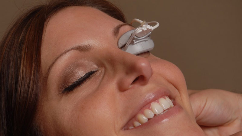
Equinox Low Level Light Therapy (LLLT)
Do You Suffer From Dry Eye?
Carolina Eye Care is now offering Marco Equinox Low Level Light Therapy (LLLT), the latest in dry eye treatment technology.
- Comfortable, non-invasive procedure
- 15 minutes per session
- No recovery time needed
- For use on adults and children
It’s quick and painless! Simply lay back with the Equinox mask on and relax as the warm light clears your glands. LLLT may also help decrease the appearance of facial wrinkles, acne and rosacea.
Corneal Topography
Corneal topography is a computer assisted diagnostic tool that creates a three-dimensional map of the surface curvature of the cornea. The three-dimensional map is a valuable aid to the examining optometrist and can assist in the diagnosis and treatment of a number of conditions; in planning cataract surgery and intraocular lens (IOL) implantation (plano or toric IOLs); in planning refractive surgery such as LASIK, and evaluating its results; or in assessing the fit of contact lenses.
Optomap Ultra-Widefield Retinal Image
The optomap ultra-widefield retinal image is a unique technology that captures more than 80% of your retina in one panoramic image while traditional imaging methods typically only show 15% of your retina at one time.
The benefits of having an optomap ultra-widefield retinal image taken are:
- Optomap facilitates early protection from vision impairment or blindness
- Early detection of life-threatening diseases like cancer, stroke, and cardiovascular disease
The unique optomap ultra-widefield view helps your eye care practitioner detect early signs of retinal disease more effectively and efficiently than with traditional eye exams